
Confession. I get my hands on every single Neuron journal I possibly can. Yep, bona fide brain geek here. Why? Because after working with kids for 40 years, I still love to watch their faces light up with that "ah ha!" moment when I explain WHAT is going on in there that makes them dyslexic or have Attention-Deficit/Hyperactivity Disorder and yes, even autism. I've learned a lot from these kids. I've learned that it makes a very, very big difference to them whether it's their "neurology" or their "personhood". If I can get them to understand their brains, then it becomes a "me versus my brain" and they take on just about any strategy I present to them in order for them to "wrestle" their brains into "behaving".
Not only do the "connections" have to work (the hardware), but so do the "fluids" or the neurotransmitters such as dopamine (motivation and satisfaction), serotonin (nature's anti-depressant), acetylcholine (sustains attention, helps with sleep, is involved in depression and is associated with Alzheimer's)..you get the picture. They're important!!!
We've learned so very much about "what" goes on in many different kinds of brains, but most recently, we've discovered the "why" behind many of the behaviors in those persons with autism. Let's take it point-by-point for the "big ticket items":
- Mirror neurons: What? These are the neurons, the brain cells, that allow you to imitate. If you have trouble imitating, you lose a major learning source. I've often wondered about those babies I see in high chairs being fed. I wonder if the babies who are watching the spoon instead of Momma turn out to have some type of "social interaction disorder". Watch someone feeding a baby; they just can't help themselves. The reason they open their mouths as they guide the spoon toward baby is because baby is watching THEM and not the spoon!!! Ta-da! Mirror neurons at work right then and there; gotcha'! Imitation is very much associated with empathy development.
- Frontal lobe: Yes, this is the part of the brain in the front! It's a really big lobe because it is the last area of the brain to develop and has many, many important roles. The pre-frontal cortex (directly behind the forehead) is intensely involved in ADHD symptoms. The frontal lobe in a person with autism is greatly enlarged due to excess brain matter and results in highly compromised executive functioning (the BIG STUFF: shifting gears, monitoring behavior and thinking; using working memory, using social judgment and forethought, planning and organizing as well as inhibiting inappropriate behavior and comments).
- Corpus callosum: (no, it's not in Texas!): It's part of the brain that connects the left and right hemispheres...a big ol' thick, flat bundle of neural fibers that facilitate communication. In the autistic brain, this part is actually undersized resulting in poor coordination of various regions of the brain and jams the messages to the sympathetic nervous system. The sympathetic nervous system has a primary role in initiating the "flight or fight" response. It is constantly active in maintaining a balance in the systems. You can see the "unreasonable" levels of fear and panic in an autistic person even when they are asked to eat a different brand of pizza. That behavior is the manifestation of this neurological deficit. Exhausting.
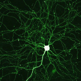
Wow! A thought or behavior in action! The axon is the brightest "string" coming out of the neuronal body. It sends messages out to the body. Cool!
- Amygdala: This part of the brain is small, but EXTREMELY powerful. It is the seat of our emotions and plays a major role in reward and yes, addiction. For those with autism, it is enlarged. In the autistic brain, the "threat" system of the amygdala is activated when looking at faces. It makes sense, doesn't it, that eye contact and looking you in the face when speaking is so painful for those with autism?
- Hippocampus: This is a Latin word for seahorse (not a hippopotamus) and describes the shape of the organ (actually there are two "facing" each other) in the brain that is responsible for memory storage. Ever wonder "why" those with autism can remember "odd information in detail years later"? It's because the hippocampus is about 10% larger, yes, 10%, then the "neurotypical" brain (we avoid using the term "normal" because it really doesn't mean much!). This enlarged hippocampus is a deficit because the person with autism relies on memory to interpret situations that most people process elsewhere in their brains.
- Cerebrum: This is largest part of the brain and contains several critical brain structures including the cerebral cortex (you think with this), the basal ganglia (you operate your motor skills with this) and the limbic system (your feelings/memory/learning). It basically overlays most of the rest of the brain. The issue here is that there is way too much white matter in this area and it interferes with motor coordination, motor planning and anticipating events. Obviously, not being able to anticipate "what's next" in a sequence of exchanges is a social liability. There's so much activity that the autistic brain is overwhelmed frequently. Again, exhausting.
White matter is a component of the central nervous system that contains glial cells and myelinated axons that send signals from one region of the cerebrum through the lower brain centers. White matter affects how the brain learns and functions. Glial cells are the "handmaidens" of neurons by keeping them in place, keeping them well-fed, keeping them away from each other and protecting them from pathogens and getting rid of dead neurons. Fun Fact: On autopsy, Einstein's brain had a prolific amount of glial cells! He had a super-clean, highly efficient brain. Not all axons (nerve fibers extending away from the neuron) are myelinated. The myelin sheath is developed by the glial cells and speeds up the electrical impulses. Multiple sclerosis is a disease of the myelinating sheath.
Even when you are in a deep sleep, your brain is always working; your mind is always active. Just look at this energy!
Now that you know the struggles of the autistic brain, you can more readily understand those persons with autism. Their lives are very challenging, even if they are high functioning autistic and the "brain facts" tell us why this is so. Be patient, be creative and most of all, be grateful.
Just do the best you can, Claudia
Join me on Facebook at Dr. Claudia McCulloch
At drclaudia.net, click on the "Ask Me" button and submit a question.
Just do the best you can, Claudia
Join me on Facebook at Dr. Claudia McCulloch
At drclaudia.net, click on the "Ask Me" button and submit a question.